Dear Reader,
The following pages of information are presented to guide you towards a better understanding of anatomy, physiology and the biomechanics of the human body. Presented is only a fraction of material available and will hopefully serve as an appetizer for your knowledge seeking.
Don Myren, D.C.
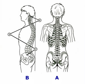
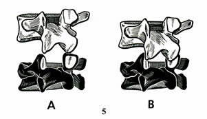
When viewed from the back, (Figure A), the vertebral column as a whole is straight. In some people, however, there may be a lateral curvature that is referred to as “Scoliosis” (That’s another topic all together). On the other hand, viewed from the side, (Figure B), the vertebral column has four curvatures.
- The sacral curve, which is fixed in adults.
- The lumbar curve, develops as a toddler begins to stand and walk.
- The thoracic curve, the largest spinal curve with 12 vertebra and 12 pair of corresponding ribs.
- The cervical curve, 7 vertebra, arguably the most influential of the spinal curves.
The vertebral column, as a whole, works much like a spring in the way it absorbs forces applied to it, specifically gravity.
LEGEND:
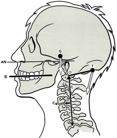
AN = Auriculo-Nasal plane
B = Bite Plate
C = Gravitational line through neck (Cervical Spine)
F = Force produced by muscles
G = Gravity, the force produced by the head’s weight
O = Fulcrum of the head movements
P = Line of perpendicularity
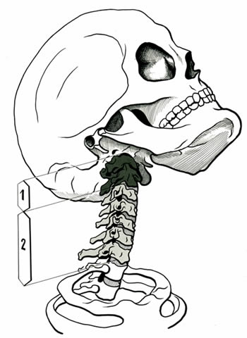
These are the deepest of the vertebral muscles. When these muscles contract (tighten) symmetrically they flatten the cervical curve and flex the neck (bend the head forward).
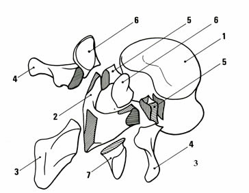
- Kidney shaped vertebral body
- Two laminae
- Spinous process
- Two transverse process
- Two Pedicle(s)
- Two superior (upper) articular process
- Two inferior (lower) articular process
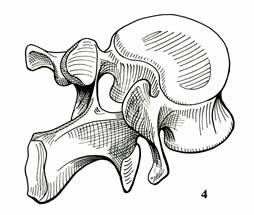
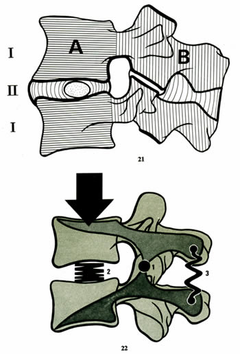
When viewed from the side (Image-21), the functional components of the column demonstrate the “pivotal” relationship of the front portion (A) and the rear portion (B) of the vertebral unit. The disc between the vertebral bodies (II) acts in effect similar to a spring or “shock absorber” (Image 22).
The “disc” is comprised of 2 parts: The annulus fibrosus is the set of circular rings of fibers which surround the centrally located jelly like material, called the nucleus pulposus.
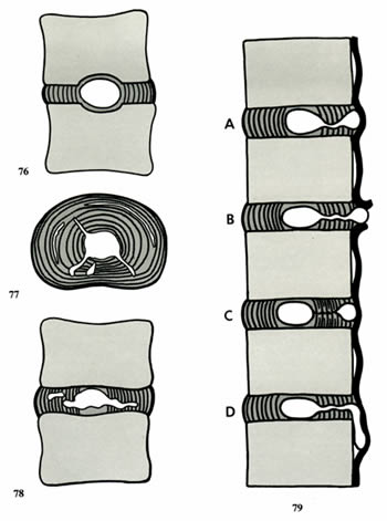
When the disc is compressed, the nucleus “jelly” can be forced into various directions.
Figure 76: Shows the nucleus forced into the bony portion if the vertebral bodies.
Figure 77: As shown from the top looking down on the disc, demonstrates the migration of the nucleus “jelly” through cracks in the annulus fiber rings.
Figure 78: Shows the nucleus material being forced into the fibrous rings, (annulus fibrosus), as viewed from the side.
This next diagram, Figure 79, demonstrates the various disc nucleus prolapse occurrences. For anatomical orientation to this diagram it is important to remember that the nucleus of the disc, the jelly, can go any of 360° of direction. However, it is when the nucleus is forced into the spinal cord and exiting spinal nerve areas that the most severe effects are felt, resulting in nerve dysfunction (pain, numbness, tingling, muscle weakness, etc.)
Figure A, The nucleus jelly has made it through the annulus fibers and is held back by a large band of ligamentous fibers that hold the vertebrae together. In this case it is still possible to bring the nuculus back into the fibers using “decompressive traction.” Go to “Spinal Decompression.”
Figure B, The nuclear jelly breaks through the final ligament fibers and is called a free floating fragment of nucleus. Depending upon the location of this “free fragment,” this can be difficult to treat non-surgically. In medical lingo, this is sometimes referred to as “sequestered piece” of nucleus.
Figure C, Here the nuclear jelly had retreated to its normal position. The annulus fibers have reattached and a portion if the nucleus has been isolated.
Figure D, In some cases the nuclear jelly can migrate underneath the ligament fibers and create problems at multiple levels. I hope these illustrations help you to visualize what can happen and the complexity of the conditions of Disc Prolapse. Remember the disc itself is a moving living structure. In as much, it can be managed if things are not left to become too severe.
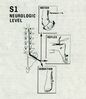
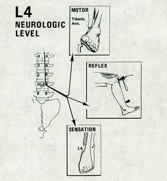
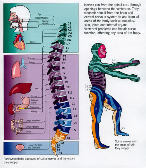
The sacrum is part of the pelvis. The first nerve exiting the sacrum is S1. This is its nerve pathway and areas of innervation. Disc problems in the lower back often affect the level below due to the anatomical position of the spinal nerves.
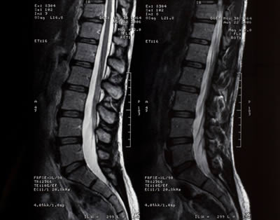
This is an image created by a computer and the introduction of magnetic fields upon the lumbar spine. This process is called an MRI Magnetic Resonance Imaging. This is the best way to visual soft (any non bone) tissues. Unfortunately, it is a bit expensive and many times reserved for pre surgical situations.
Click Here to contact us if you would like more information or to make an appointment